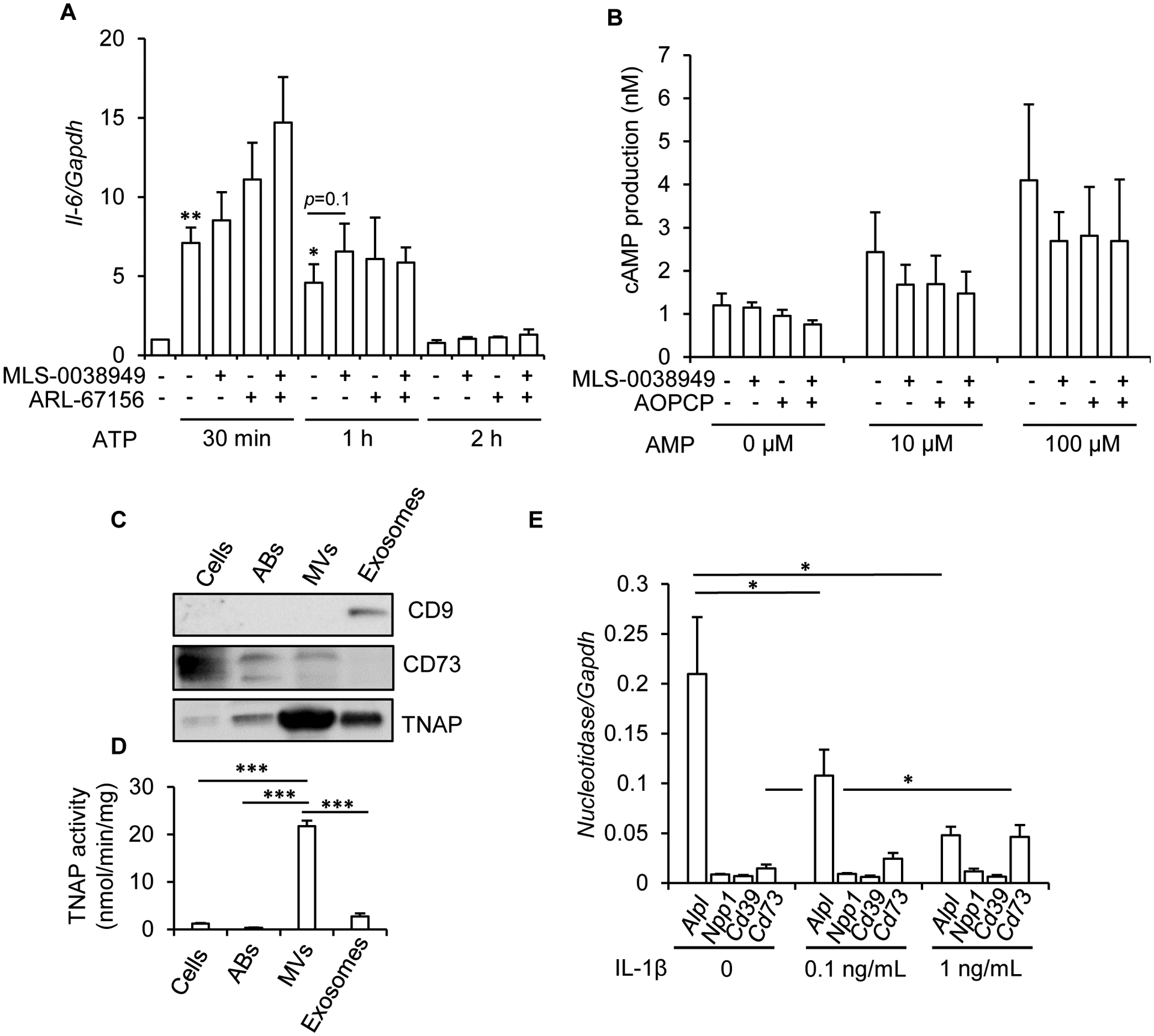
Figure 4: absence of anti-inflammatory effects of TNAP in hypertrophic chondrocytes.

Mouse primary chondrocytes were differentiated into hypertrophic chondrocytes for 16 days as detailed in the Materials and methods section. A: effect of MLS-0038949 and/or ARL-67156 on ATP-stimulated Il-6 expression (n=8). To eliminate the inter-experimental variations in Il-6 levels, Il-6/Gapdh ratio in untreated cells was set to 1. B: effect of MLS-0038949 and/or AOPCP on AMP-stimulated intracellular cAMP production (n=4). C: western-blot analysis of TNAP, CD73, and CD9 in hypertrophic chondrocyte cells, and in apoptotic bodies (ABs), MVs and exosomes released by these cells (n=3, a representative result is shown; the same quantity of protein was used in all conditions). D: TNAP activity in hypertrophic chondrocyte cells, and in ABs, MVs and exosomes released by these cells (n=4). E: effect of a 24-h treatment with IL-1β on nucleotidase levels, as determined by RT-qPCR (n=4). * indicates a statistical difference with p<0.05; ** a difference with p<0.01, and *** a difference with p<0.001.
