
Figure 3: Vagal ganglion neurons transmit sugar signals to the brain.
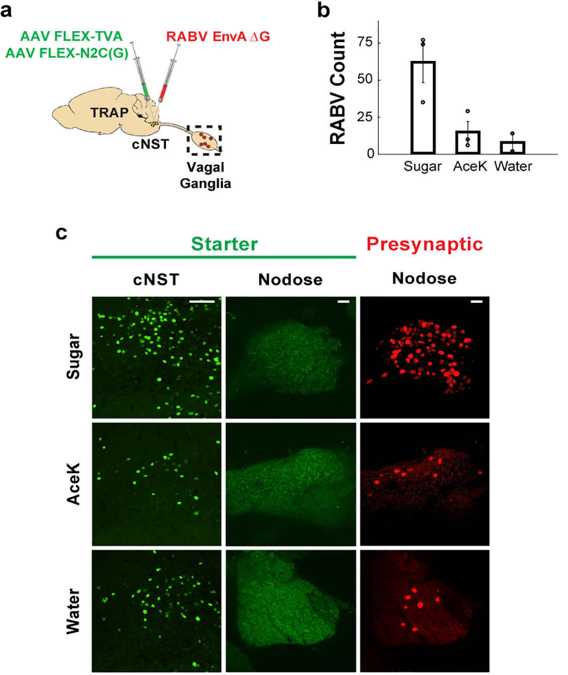
a, Strategy for targeting a red-fluorescently labelled retrograde transsynaptic rabies reporter (RABV-dsRed)28,31 to the cNST. Sugar-TRAP neurons in the cNST (defined as “starter”)30,31 were infected with the RABV-dsRed virus; the monosynaptic inputs of the sugar-activated cNST neurons are revealed by the retrogradely transsynaptic transfer of the RABV-dsRed virus. b, Quantification of retrogradely-labelled RABV-dsRed neurons in the nodose ganglion. Sugar- versus AceK-TRAPing (n = 3 animals), ANOVA Tukey’s HSD post hoc test, p = 0.0449. We also performed control TRAPing with water (n = 2 animals); Sugar- versus water-TRAPing, ANOVA Tukey’s HSD post hoc test, p = 0.0407; AceK versus water-TRAPing, p = 0.9. Values are mean ± s.e.m. c, Sugar-TRAPed cNST neurons (“starter”, green) receive monosynaptic input from vagal neurons (RABV, red). Note the absence of starter cells in the nodose, confirming that the RABV (red) cells represent retrogradely-labelled neurons30,31. Scale bars, 100 μm.