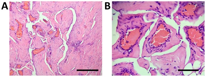
Figure 4.
Histological images of the excised cervical vertebrae indicate proliferative fibrous tissues and numerous thin-walled blood vessels in the bone cavity without evidence of cellular atypia and osteogenesis reactions. (A) Magnification, x100, scale bar, 200 µm; (B) magnification, x400, scale bar, of 50 µm; Hematoxylin and eosin staining.

