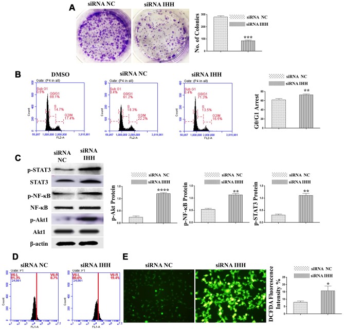
Figure 3.
Silencing of IHH induced CFU inhibition, cell cycle arrest, and aging-related signaling pathways in BMSC. (A) BMSC (n = 5) were transfected for 24hours and then incubated in 10% FBS in MEM-ALPHA medium for 12 days. Colonies were visualized after staining by 0.02% crystal violet stain. (B) BMSC (n = 5) were transfected with siRNA negative control, siRNA IHH or DMSO for 24hours. Fixed cells stained by PI and RNase A, and then analyzed by flow cytometry for cell cycle distribution (C) BMSC (n = 5) were transfected with siRNA negative control or siRNA IHH for 48hours. Akt1, p-Akt1, NF-κB, p-NF-κB, STAT3, and p-STAT3 proteins expressions were measured by Western Blot. β-actin was used as an internal control. (D) BMSC (n = 5) were transfected with siRNA negative control or siRNA IHH for 24hours then stained by DCFHDA (5 μM). The fluorescent intensity of ROS was measured by flow cytometry and immunofluorescence microscopy (E). Results are shown as mean ± SEM. *P <0.05, **P < 0.01, ***P < 0.001, ****P < 0.0001.

