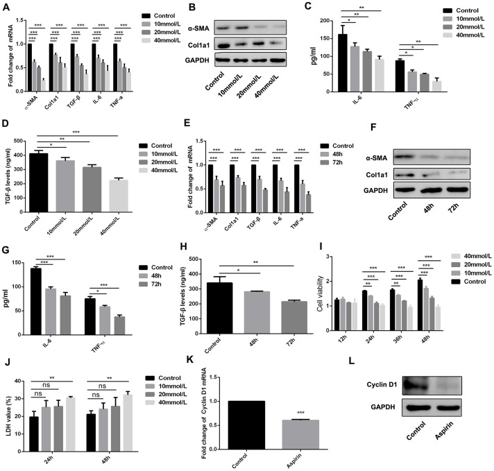
Figure 3.
Aspirin inhibited the activation and proliferation of hepatic stellate cells. (A, B) Real-time PCR and western blot was employed to examine the expression of α-SMA, collagen-a1, TGF-β, IL-6 and TNF-α in the LPS-treated HSCs with disposure of 0, 10, 20, 40mmol/L. (C, D) ELISA assay was used to examine the expression of TGF-β, IL-6 and TNF-α in the LPS-treated HSCs with disposure of 0, 10, 20, 40mmol/L. (E, F) Real-time PCR and western blot was employed to examine the expression of α-SMA, collagen-a1, TGF-β, IL-6 and TNF-α in the LPS-treated HSCs with disposure of 40mmol/L at 48 and 72h. (G, H) ELISA assay was used to examine the expression of TGF-β, IL-6 and TNF-α in the LPS-treated HSCs with disposure of 40mmol/L at 48 and 72h. (I) CCK-8 assays were performed to examine the proliferation of LPS activated-HSCs with aspirin treatment. (J) LDH assay was performed to detect the cell viability of LPS activated-HSCs with aspirin treatment. (K, L) The expression of Cyclin D1 was detected by real-time PCR and western blot. *P<0.05, **P<0.01, ***P<0.001.

