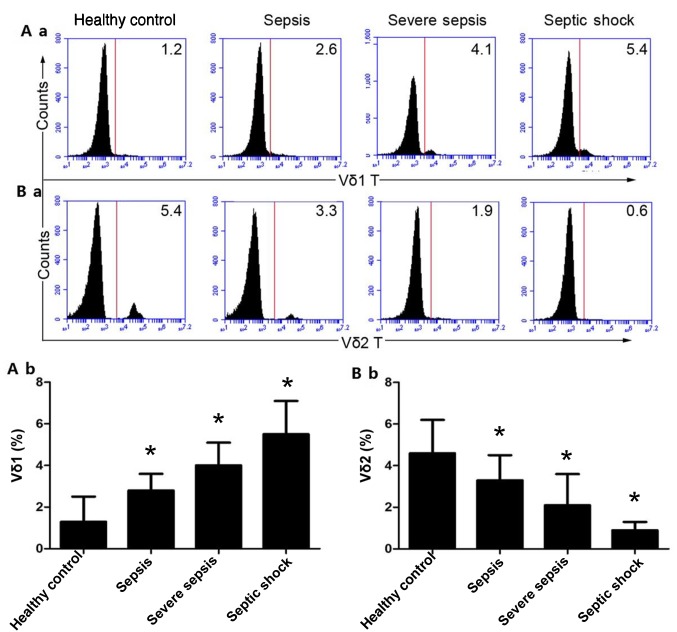
Figure 2.
Ratios of Vδ1 and Vδ2 T cells in patients with different types of sepsis. (A-a) Percentage of Vδ1 T cells was measured by flow cytometry. (A-b) Quantitative analysis of Vδ1 T cells. (B-a) Percentage of Vδ2 T cells was measured by flow cytometry. (B-b) Quantitative analysis of Vδ2 T cells. The histograms are representative examples of the data (14 patients with sepsis, 9 patients with severe sepsis, 7 patients with septic shock and 30 healthy controls). *P<0.01 vs. healthy control.

