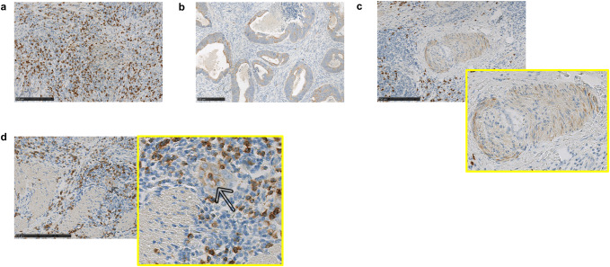
Fig. 1.
IL-36γ is expressed by a variety of cell populations in the tumor microenvironment. FFPE tumor sections were stained for IL-36γ by IHC as described in “Materials and methods” for expression of IL-36γ. Expression of this protein was observed in immune cells (a), tumor cells (b), and cells of the vasculature (c, d). Both smooth muscle (c) and endothelial cells (d) of the vasculature were observed to express IL-36γ. Bars 250 μm

