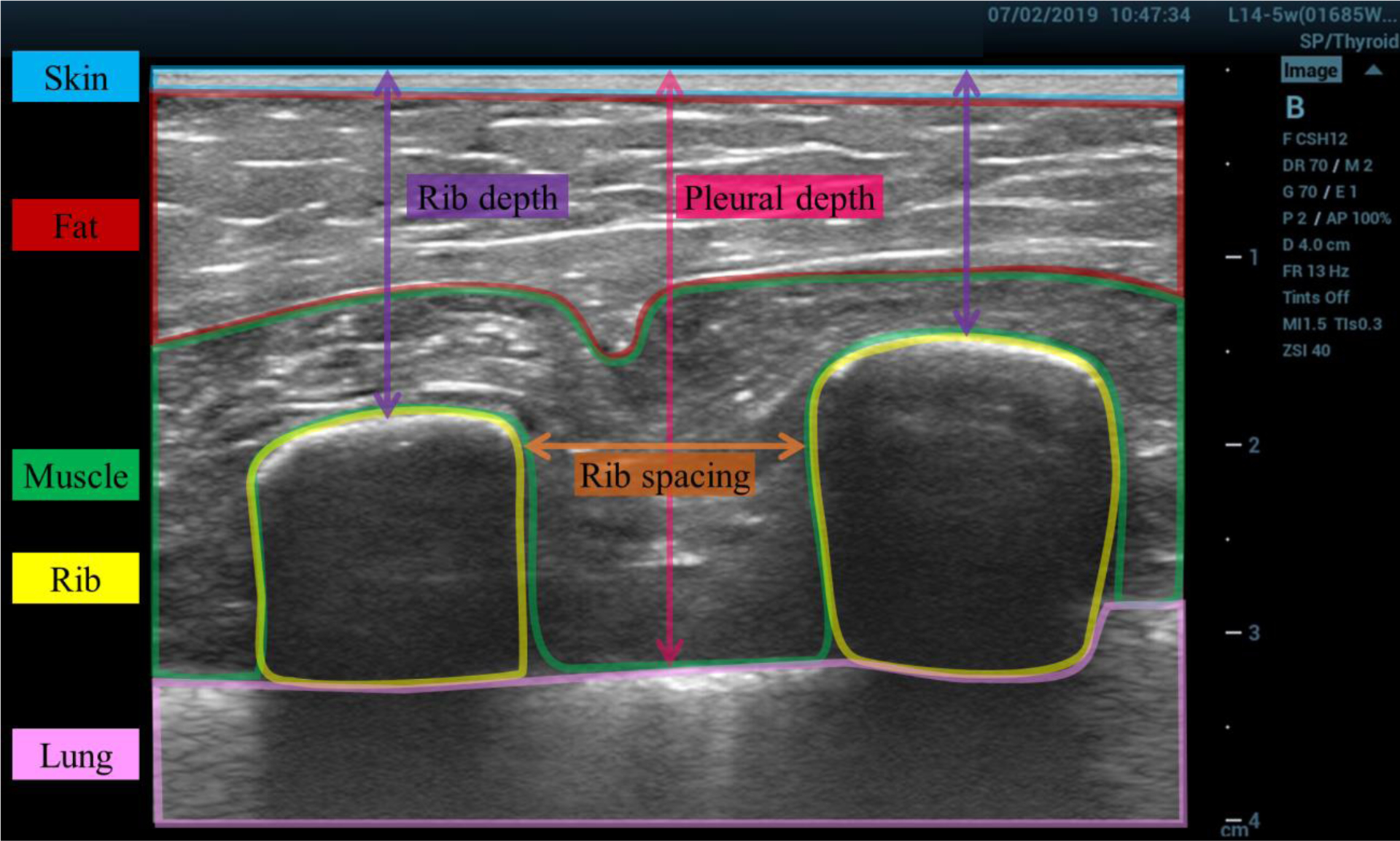
Figure 1:

Example ultrasound image of the chest wall, with labeled tissue layers. The image was taken in situ prior to excision. The approximate borders of different tissue layers are highlighted and labeled: skin (blue), fat (red), muscle (green), rib (yellow), lung (pink). The rib spacing (orange), rib depth (purple), and pleural depth (magenta) measurements are illustrated.
