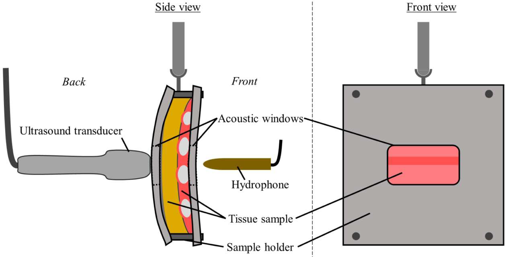
Figure 2:

The experimental setup within the water bath. The ultrasound probe, hydrophone and tissue sample in its holder are shown. The hydrophone is rigidly mounted, while the probe and tissue sample are attached to 3D micropositioning systems (not shown). Within the tissue sample, the fat, muscle, and ribs are shown in yellow, pink, and light gray respectively.
