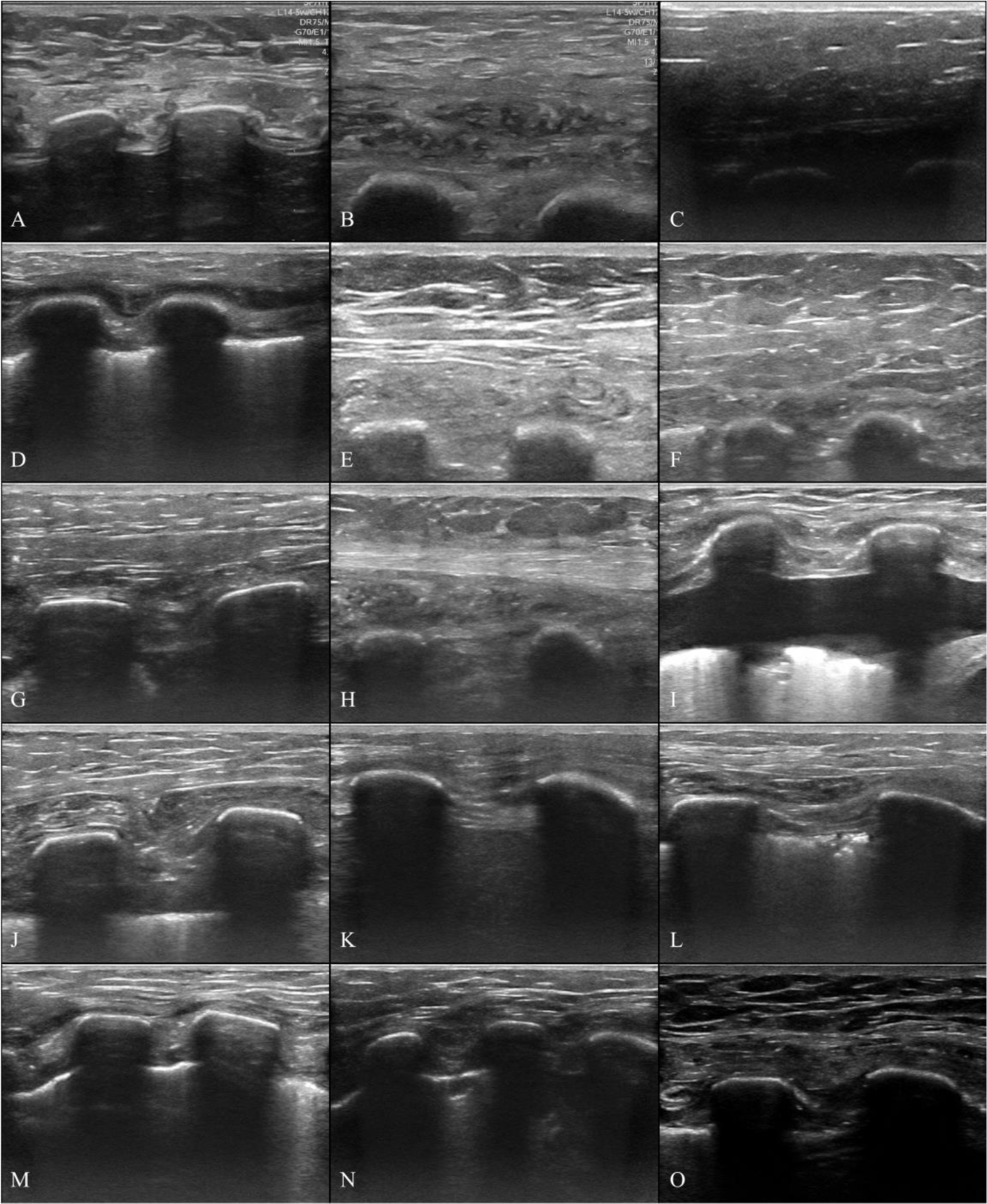
Figure 3:

In situ ultrasound images of human chest wall samples. Images were taken immediately before the samples were excised. Images are 4 cm deep and highlight differences in tissue sample morphology between donors. Each image shows a thin layer of skin at the top, subdermal soft tissue (e.g. adipose, muscle, connective tissue, etc…), and two (three for Sample N) ribs.
