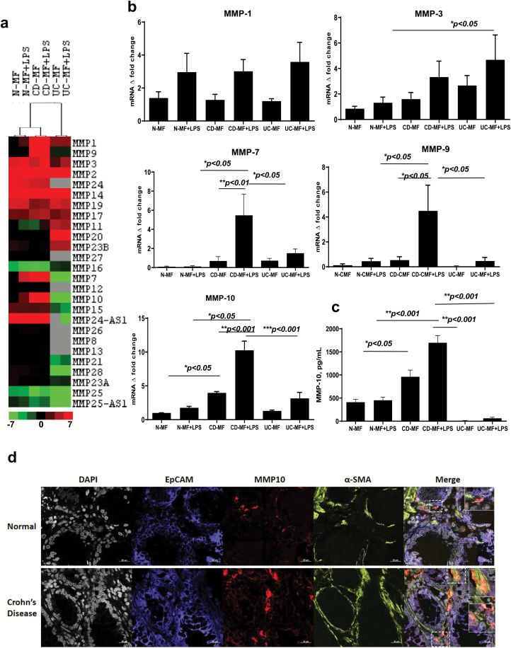
Fig. 1.
Expression of basal and LPS-inducible patterns of MMPs differs in CD-MFs when compared to N- and UC-MFs. (a) mRNA microarray analysis shows differential MMP expression in CD-MFs compared to N-MFs and UC-MFs at basal and TLR4-stimulated conditions. Sample-based hierarchical clustering was carried out as described in Methods. The color chart at the bottom of the figure shows levels of fold decrease and increase, log2 fold changes are shown. (b) Basal and LPS-inducible differential expression of MMP-7, -9 and -10 by MFs isolated from normal, CD and UC mucosa was analyzed on mRNA level using real-time PCR. (c) Basal and LPS-inducible secretion of MMP-10 by MFs isolated from CD mucosa is increased when compared to normal, and UC mucosa-derived MFs, multiplex MMP array analysis. LPS was used at concentration 1 μg ml−1, 72-h exposure. Data are shown as means ± SEM, n = 5, *P < 0.05; **P < 0.01; ***P < 0.001. (d) MMP-10 expression is increased within α-SMA+ MFs in CD intestinal mucosa when compared to the healthy controls. Confocal microscopy images of representative cross-sections from CD and normal human intestinal mucosa (n = 4 per group) are shown. MFs were detected by anti-α-SMA mAb (in green), and epithelial cells were identified with anti-EpCAM mAb (in blue). Co-localization of MMP-10 (in red) with α-SMA+ MFs results in formation of yellow-orange color on merged images. High resolution areas in the merged images are outlined in the boxes.

