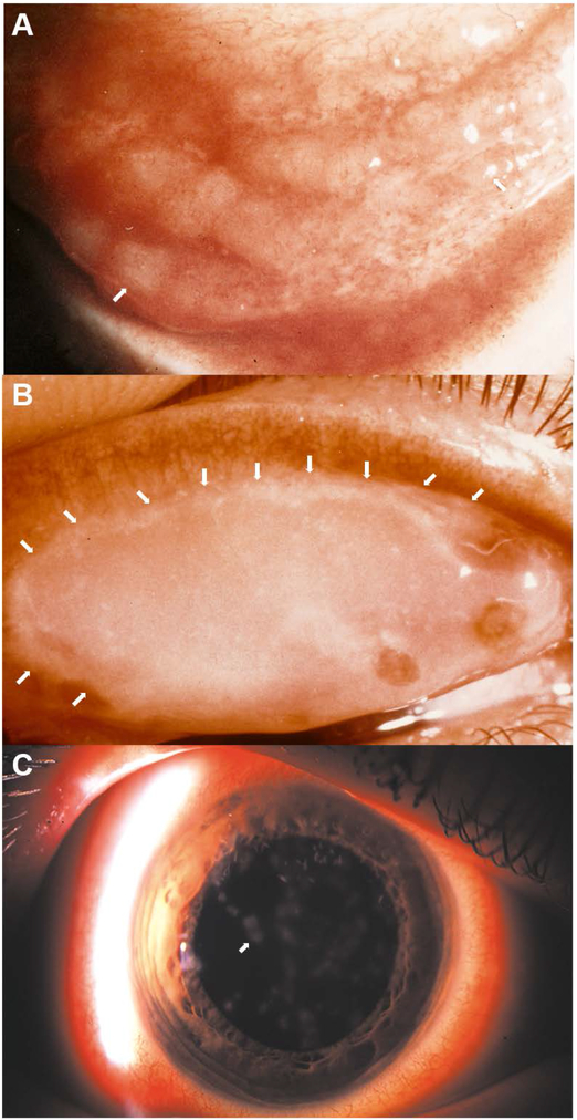
Fig. 1.
Photomicrographs of common clinical manifestations of ocular surface in epidemic keratoconjunctivitis (EKC). (A) Inferior conjunctival fornix of a patient with acute EKC shows conjunctival lymphoid hyperplasia presenting as jelly bean-shaped milky elevations of the mucosa (arrows point to two out of many follicles). (B) Superior eyelid tarsal surface with a conjunctival membrane (with visible margins of the membrane delineated by white arrows). (C) Corneal subepithelial infiltrates (arrow points to one of many such infiltrates). Image in C reproduced with permission.[264]

