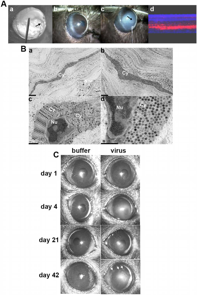
Fig. 7.
Mouse model of adenovirus keratitis. (A) Induction of the mouse model of adenovirus keratitis. a. BALB/c mouse cornea retroilluminated to show size of heat-pulled, glass micropipette needle (arrow) as compared to a 33 gauge metal needle. b. Diffuse illumination view of mouse cornea prior to, and c. during injection with HAdV-D37 or virus-free dialysis buffer using the glass needle (arrow) and a gas-powered microinjection system. The injection causes transient whitening of the corneal stroma (arrow points to tip of glass needle within the corneal stroma. d. Composite confocal image of a mouse cornea taken immediately after injection of Cy3 dye demonstrates successful intrastromal injection. (B) Thin-section electron microscopy of C57Bl/6j mouse corneal stroma at intermediate time points after intrastromal injection of HAdV-D37. Micrographs show the corneal stroma at a, 4 hours, and b-d, 8 hours after injection. Intracellular structures are labeled as follows: Cy, cytoplasm; Nu, nucleus. The inset in a shows a higher magnification of intracellular virus. All micrographs show densely packed intracellular viral arrays. Scale bars: 2 μm in a and b, and 0.5 μm in c and d, and the inset in a. Adapted with permission from Mukherjee et al.[233] (C) Representative clinical photographs of C57BL/6j mouse corneas after mock infection with dialysis buffer or 105 TCID HAdV-D37 at days shown post infection. Buffer-injected corneas remained clear at all times. Opacities in HAdV-D37 injected corneas were seen as early as 1 day after infection, and appeared to peak at 4 days. The opacities then regressed slowly but in approximately one-third of mice recurred as characteristic subepithelial infiltrates at about 6 weeks after infection (day 42, right panel, arrows). Figure adapted with permission from Chintakuntlawar et al.[232]

