
Figure 4.
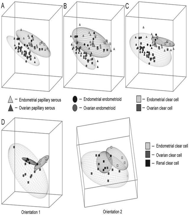
Principle component analysis (PCA) of ovarian and endometrial cancers according to histology. (A) PCA of tumors with serous histology showing two nonoverlapping elliptical regions separating endometrial (top) from ovarian (bottom) specimens. (B) PCA of tumors with endometrioid histology showing two nonoverlapping elliptical regions separating endometrial (top) from ovarian (bottom) specimens. (C) PCA of tumors with clear cell histology showing overlapping elliptical regions representing endometrial (top) and ovarian (bottom) specimens. (D) PCA of tumors according to organ of origin shows three overlapping elliptical regions among ovarian, endometrial, and renal clear cell specimens, with two different orientations (1 and 2).