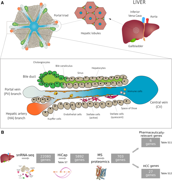
FIG. 1.
Schematic cross-sectional view of the structural organization and cell populations of a liver lobule. (A) Each lobule presents a radial structure with a central vein (CV) in the middle from which HC cords radiate toward the so called portal triad consisting of branches of the portal vein (PV) and hepatic artery (HA) and bile ducts. The sinus is delimited by HCs that are arranged back to back in cords and it is lined by specialized sinusoidal endothelial cells. Kupffer and immune cells are located in the sinusoidal lumen, while hepatic stellate cells are localized in the space of Disse. (B) Workflow representing the design of the multi-omics study. The crystal structure of the glucose transporter was obtained from PDB (ID: 4ZWB, Deng et al., 2015). HC, hepatocyte.

