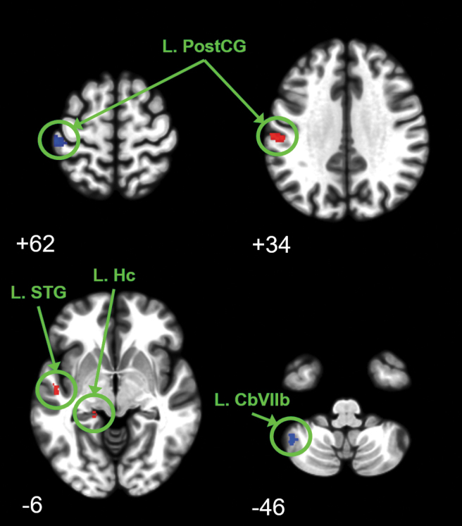
FIG. 5.

Regions of altered brain connectivity for mild traumatic brain injury (TBI) subjects over 1 year including 9 months of growth hormone (GH) replacement. Significant change in connectivity was determined with flexible factorial analysis using peak-level familywise error (FWE) corrected p < 0.05. Clusters ≥8 voxels are reported with blue clusters depicting decreased connectivity and red clusters depicting increased connectivity. Images are depicted at Montreal Neurological Institute (MNI) oriented axial slice number. Connectivity was decreased between the right frontal eye field and the left post-central gyrus (upper left image) whereas connectivity increased between the left thalamus and the left post-central gyrus (upper right image). Connectivity was increased between the left post-central gyrus and the left superior temporal gyrus and left hippocampus (lower left image). Connectivity was decreased between the posterior cingulate and the left cerebellar (Cb) lobule VIIb (lower right image).
