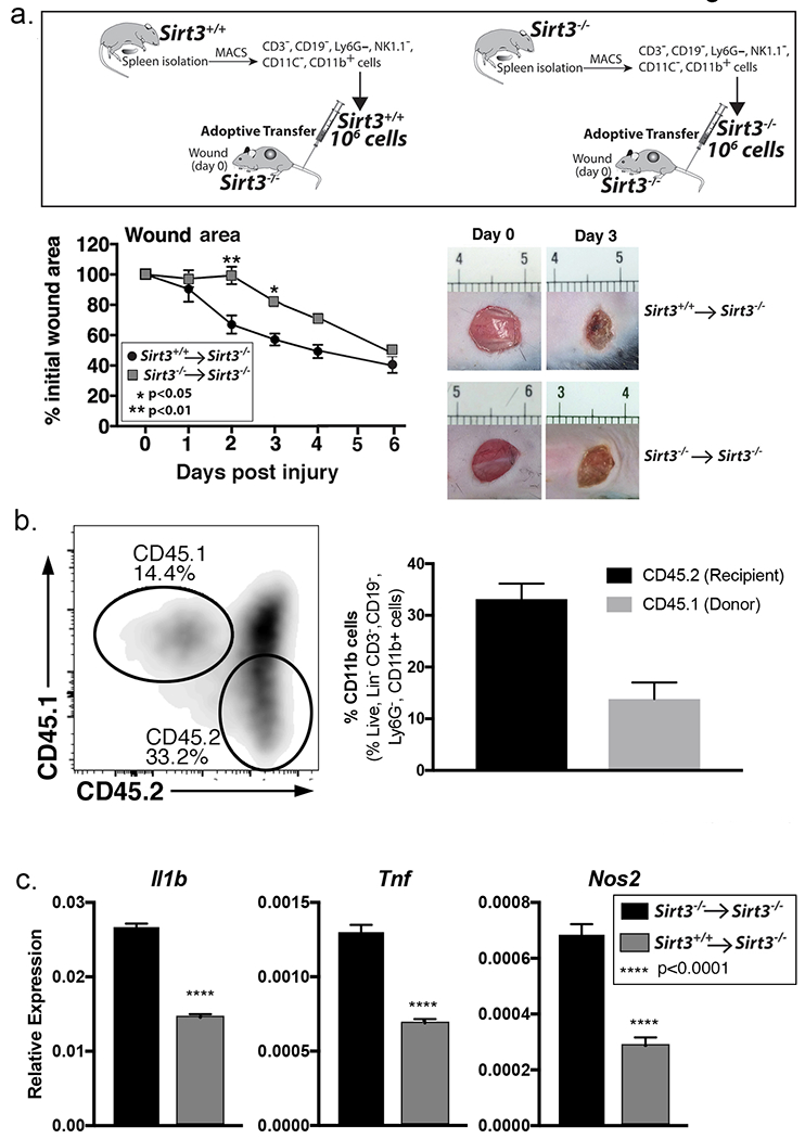
Figure 2. Adoptive transfer of Sirt3-expressing macrophages decreases inflammation and improves wound healing in Sirt3-deficient mice.

A: Monocyte/macrophage (CD11b+CD3−CD11c−CD19−Ly6G−NK1.1−) single cell suspensions were isolated from Sirt3−/− and Sirt3+/+ spleens by cell sorting. 1 × 10⁶ cells were injected intravenously into wounded Sirt3−/− mice. Wound closure was measured using ImageJ software (n=5 mice, repeated 1x). B: Monocyte/macrophage (CD11b+CD3−CD11c−CD19−Ly6G−NK1.1−) single cell suspensions were isolated from B6.SJL-PtprcaPepcb/BoyJ spleens by cell sorting. 1 × 10⁶ cells were injected intravenously into wounded Sirt3−/− mice. Wounds were harvested and processed for CD11b+CD3− CD19−Ly6G− 3 days post-injury and analyzed for CD45.1 and CD45.2 surface marker expression by flow cytometry. Bar chart shows relative levels of CD45.1 and CD45.2 as a percent of CD11b+CD3− CD19−Ly6G− cells (n=3 mice). C: Monocyte/macrophage (CD11b+CD3−CD11c−CD19−Ly6G−NK1.1−) single cell suspensions were isolated from Sirt3+/+ or Sirt3−/− spleens by cell sorting. 1 × 10⁶ cells were injected intravenously into Sirt3−/− mice and wounded. Wounds were harvested and processed for CD11b+CD3− CD19−Ly6G− 3 days post-injury. Relative gene expression of Il1b, Tnfa, and Nos2 was measured (n=6 mice). Data are presented as mean ± SEM. Data were analyzed for normality and 2-test Student t-test was performed. For data with multiple comparisons, ANOVA followed by Newman-Keuls multiple comparisons test was performed.
