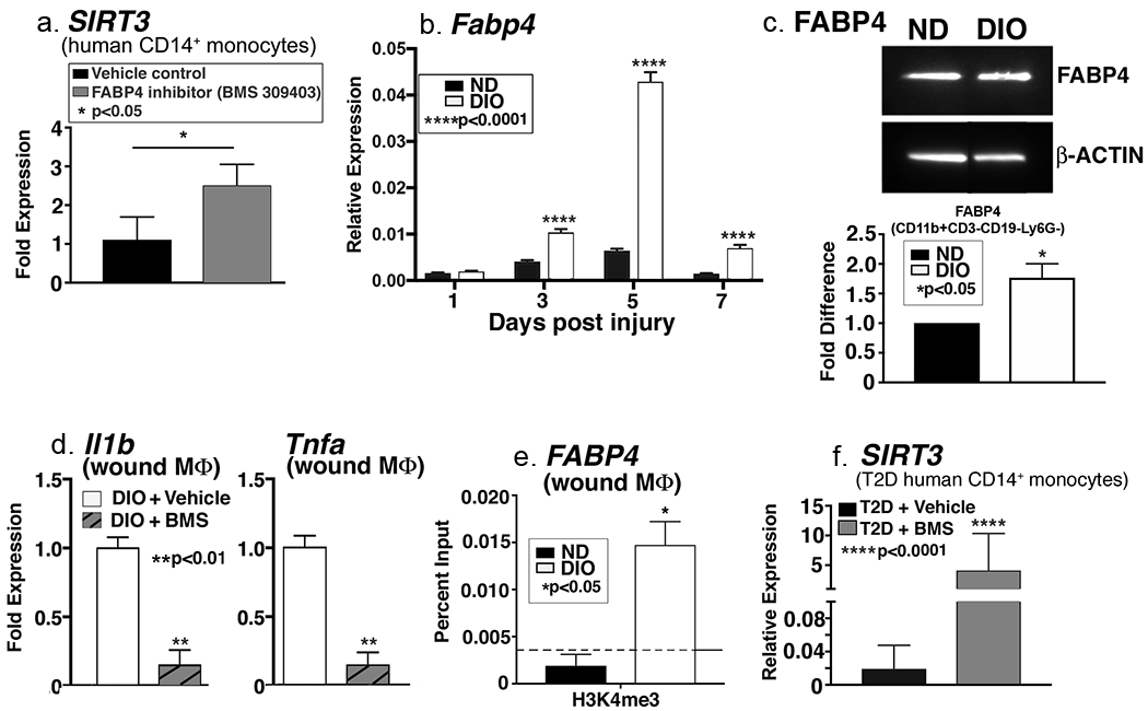
Figure 4. FABP4 regulates SIRT3 in human monocytes.

A: Human peripheral blood monocytes (CD14+) were isolated and incubated ex vivo with FABP4 inhibitor (BMS309403) or vehicle control for 6 hours. Related SIRT3 gene expression was quantified and is shown as fold expression compared to vehicle-treated (n=3, repeated 1x in triplicate). B: Wound macrophages (CD11b+CD3−CD19−Ly6G−) were isolated from DIO mice and controls on days 1, 3, 5, and 7 post-injury by cell sorting. Relative Fabp4 gene expression is shown (n=4 mice/time point, repeated 2x). C: FABP4 protein expression was assessed using Western Blot analysis (n=4 mice, repeated 2x). D: DIO wound macrophages (CD11b+CD3−CD19−Ly6G−) were isolated and incubated ex vivo for 6h with an FABP4 inhibitor (BMS309403) or vehicle control. Il1b and Tnfa gene expression is shown as fold change compared to vehicle-treated (n=14 mice, repeated 2x). E: DIO and control wound macrophages (CD11b+CD3−CD19−Ly6G−) were isolated by cell sorting on day 3. ChIP analysis was performed for H3K4me3 at the Fabp4 promoter (n=12 mice, repeated 2x). F. Human peripheral blood monocytes (CD14+) were isolated from diabetic patients and incubated ex vivo with FABP4 inhibitor (BMS309403) or vehicle control for 6 hours. Sirt3 gene expression was quantified (n=3, repeated 1x). Data are presented as mean ± SEM. Data were analyzed for normality and 2-test Student t-test was performed. For data with multiple comparisons, ANOVA followed by Newman-Keuls multiple comparisons test was performed.
