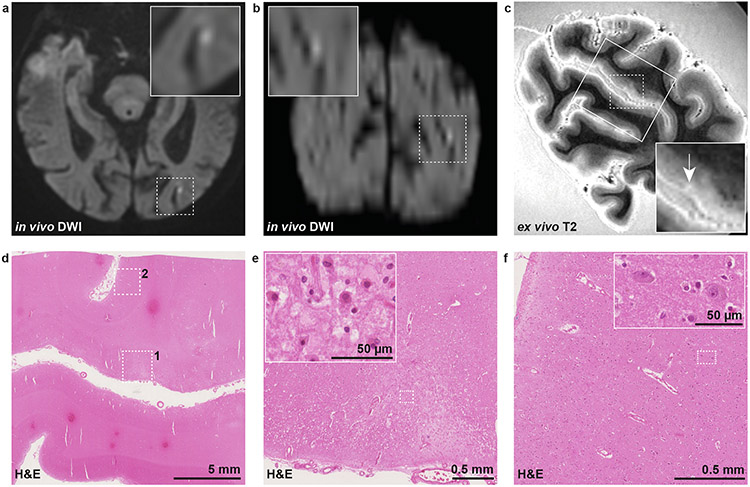
Fig. 1. Histopathological verification of an in vivo DWI+ lesion in CAA.
An in vivo cortical DWI+ lesion was detected in the left occipital lobe of a case with CAA on clinical MRI 7 days before death (a: axial view, b: coronal view). Five coronal slabs covering this region were subjected to high-resolution 7 T ex vivo T2 to guide tissue sampling as part of a previous study [34], and a T2 hyperintense lesion which may correspond to the in vivo DWI+ lesion was identified (c, white arrow). A small tissue block (outlined on T2 with rectangle) was cut and subjected to H&E staining (d). On H&E, a focal area of tissue pallor (dotted square 1) was identified with evidence of eosinophilic necrosis (inset in e), indicative of recent ischemia. In a control cortical area nearby (dotted square 2 in d) no evidence of eosinophilic necrosis was seen (f). Note that the chance for a false-positive finding was high in this case, owing to the large difference in resolution between in vivo and ex vivo MRI. DWI = diffusion-weighted imaging. H&E = hematoxylin & eosin.

