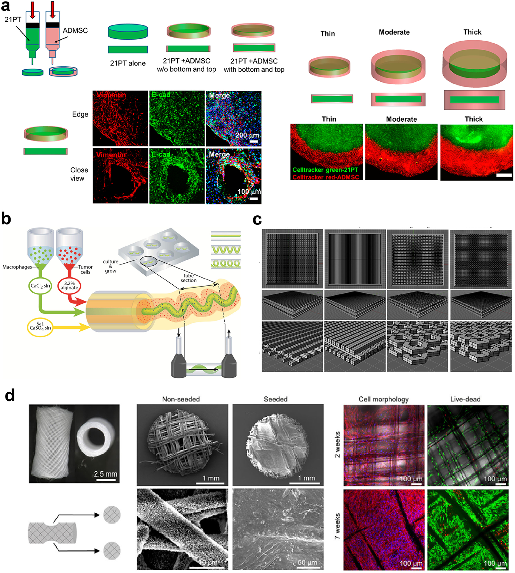
Figure 4.

Three dimensional printing is an efficient method in modeling the TME, as it enables spatial and morphological control on the printed materials. Multiple print heads enable (a) bioprinting of the tumor cells in the center and the mesenchymal stem cells (MSCs) in the outer region to mimic the TME (reproduced with permission from [255], Copyright 2018 American Chemical Society) or (b) co-bioprinting of the immune cells and the tumor cells, the immune cells being printed as blood vessel-like channels passing through the tumor (reproduced with permission from [260], Copyright 2015 John Wiley and Sons). (c) Scaffolds can be printed in various shapes thanks to computer aided design (CAD). Reproduced with permission from [261]. Copyright 2016 Elsevier Science. (d) 3D printing can also be used to recreate the metastasis process. Here, breast-to-bone metastasis of tumor cells is simulated using the 3D printed humanized bone. Reproduced with permission from [263]. Copyright 2014 The Company of Biologists Ltd. Creative Commons Attribution 3.0 License.
