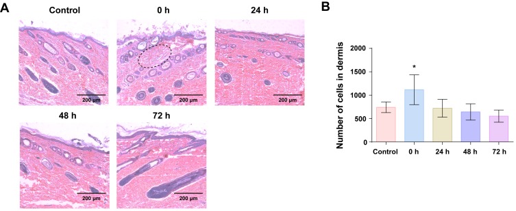
Figure 7.
Histopathological analysis of guinea pig skins after the combined use of hucMSC-Exos (150 μL, 1 mg/mL) and SHSs over 72 h. (A) Histopathological images of the skins. Dashed circle: inflammatory infiltration. (B) Numbers of cells in a certain section area (450 μm × 150 μm) of dermis. All data were expressed as mean ± SD (n = 3). *p < 0.05, versus the control group.

