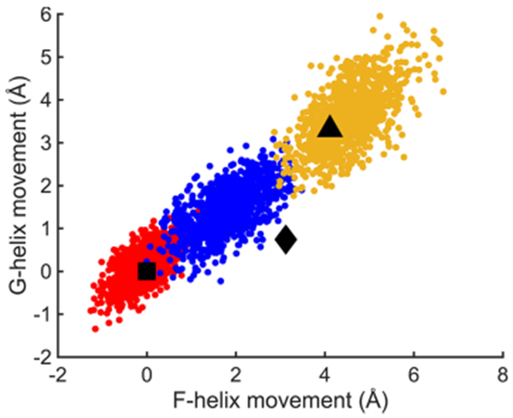
Figure 3.

Movements of F and G helices derived from MD trajectory snapshots in three ferric P450cam complexes, camphor/P450cam (red), CN−/camphor/P450cam/Pdx (blue), and camphor/P450cam/Pdx (yellow) relative to the camphor-bound state (PDB ID 3l63). Square, diamond, and triangle symbols represent the movements of F and G helices observed in crystal structures of P450cam in the closed camphor-bound (PDB ID 3l63), intermediate tethered adamantane (camphor analogue)-bound (PDB ID 1re9), and open camphor/Pdx-bound (PDB ID 4jx1) P450cam complexes.
