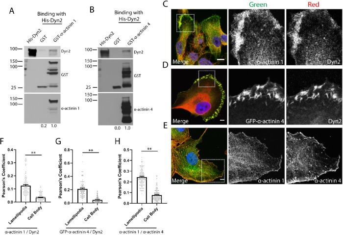
FIGURE 1:
Dyn2 binds to α-actinin 1 and 4 and colocalizes in lamellipodia of migrating tumor cells. (A, B) GST pull down of full-length GST–α-actinin 1 (A) or GST–α-actinin 4 (B) was performed to test direct binding to full-length His-Dyn2. GST proteins were blotted with α-actinin 1 and α-actinin 4 antibodies to validate their identity. n = 3 independent experiments, densitometry was performed to measure binding, and the relative average binding values are listed below each lane. (C–E) Immunofluorescence of α-actinin 1/4 and Dyn2 in PANC-1 cells reveals these proteins colocalize in lamellipodia in PDAC cells. The region highlighted in the Merge image is shown in the individual channel insets. Scale bars: 10 μm. (F–H) Pearson’s coefficients were measured to quantify where α-actinin 1/4 and Dyn2 colocalize in tumor cells. For each cell analyzed, the colocalization between indicated proteins was quantified in the lamellipodia and in the cell body. Graphed data represent the mean ± SEM, and data points represent individual cells. Between 70 and 101 cells were quantified across three independent experiments. Scale bars: 10 μm. Student’s t test was used to measure statistical significance. ** indicates p < 0.01.

