FIGURE 3:
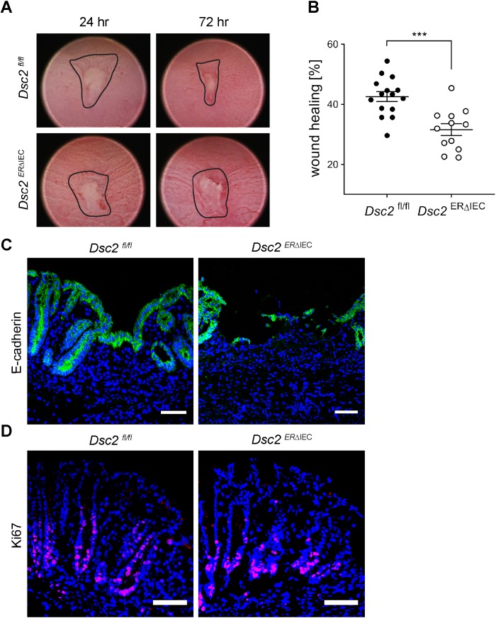
Loss of Dsc2 in IEC resulted in impaired colonic mucosal healing after biopsy-induced wound injury. (A) Representative endoscopic images of biopsy-induced colonic wounds at 24 and 72 h postinjury in Dsc2ERΔIEC and Dsc2fl/fl mice. (B) Digital measurement of wound surface at 24 and 72 h postwounding revealed significant impairment of wound healing in absence of Dsc2 on IEC. Scale bar: 50 μm. Dots represent the mean value within three to five wounds from individual mice. Data are combined values of two independent experiments with 12–15 mice per group and are expressed as mean ± SEM. Significance is determined by two-tailed Student’s t test. ***p ≤ 0.001. (C) IF images of frozen sections of wound beds at 72 h postinjury stained with the epithelial marker E-cadherin (green) and DAPI (blue) showing a dramatic impairment of wound closure in Dsc2ERΔIEC mice. Dsc2fl/fl mice present a layer of wound-associated epithelial cells that covers the wound that is absent in Dsc2ERΔIEC mice. (D) Ki67 staining (magenta) and DAPI counterstain (blue) of frozen sections of crypts immediately adjacent to wound beds at 72 h postinjury revealed similar proliferation rate between Dsc2ERΔIEC and Dsc2fl/fl mice. C and D show representative IF images of five mice per group. Scale bar: 100 μm.

