FIGURE 4:
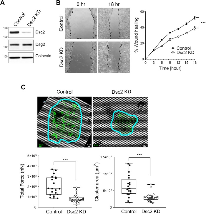
Loss of Dsc2 results in impaired wound closure and cell–matrix adhesion in vitro. SKCO15 cells were transduced with Dsc2-specific shRNA (Dsc2 KD) or with a nontargeting shRNA (control). (A) KD of Dsc2 expression was confirmed in SKCO15 by Western blot using anti-Dsc2 antibodies and anti-calnexin as loading control. No major changes were detected in the expression of Dsg2 in the absence of Dsc2. (B) Monolayers of SKCO15 cells were scratch-wounded and monitored for wound closure. Dsc2 KD cells showed significant delayed wound repair compared with control cells. Representative images of wounds at 0 and 18 h. Graph is representative of two independent experiments with three replicates per group and with two independently transduced SKCO15 cell culture. Data are mean ± SEM. Significance is determined by two-way ANOVA. ***p ≤ 0.001. (C) Measurement of traction forces as well as cluster area within SKCO15 cell KD for Dsc2 (Dsc2 KD) or control cells by using fibronectin-coated microfabricated postarray detectors (mPADs) (see Materials and Methods). Representative images show posts (white) with the cell cluster outlined in blue and force vectors (green) calculated from post deflections. Total traction force represents the sum of the magnitudes of the force vectors for each cell cluster. Data consists of 20 cell clusters per condition and are representative of two independent experiments with two independently transduced SKCO15 cell culture. Significance is determined by two-tailed Student’s t test. ***p ≤ 0.001. Scale bars: 5 μm. Scale for force: 10 nN.

