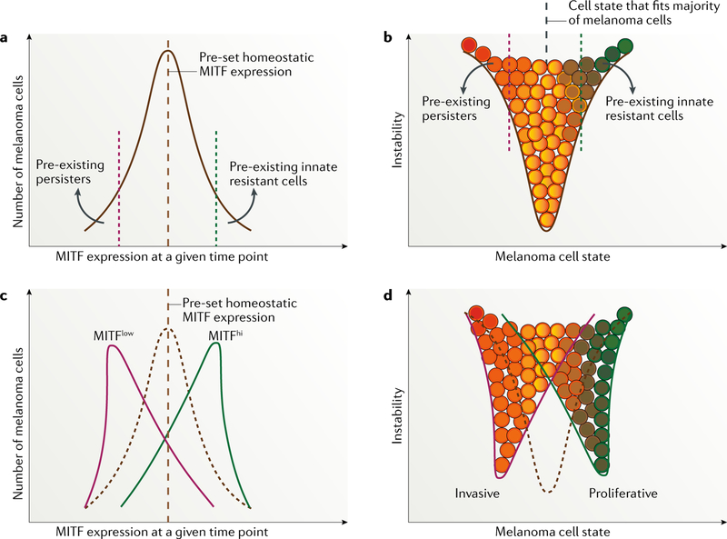
Fig. 2 |. MITF expression and phenotype switching in melanoma before and after treatment with targeted agents.
a,b | Microphthalmia-associated transcription factor (MITF) expression and melanoma cell-state pattern before exposure to targeted therapy, representing a unimodal distribution of MITF expression in the same population of melanoma cells. Of note, variations in predefined points exist between melanoma cell lines, mouse models and patient samples 72,225 owing to differences in melanoma initiation and in microenvironment selective pressure. c,d | Alterations in MITF expression and variation in cell state after starting therapy.

