FIGURE 3:
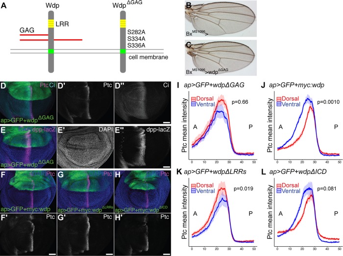
Wdp negatively regulates Hh signaling in a GAG-dependent manner. (A) A schematic drawing of WT Wdp and a mutant form of Wdp (WdpΔGAG). (B, C) A control adult wing (B) and a wing expressing UAS-wdpΔGAG with BxMS1096-GAL4 (C). (D, E) Wing discs expressing UAS-wdpΔGAG with ap-GAL4 were immunostained for Ptc, Ci (D), and dpp-lacZ (E). (F–H) Wing discs expressing UAS-3xMyc:wdp (F), UAS-3xMyc:wdpΔLRRs (G), and UAS-3xMyc:wdpΔICD (H) with ap-Gal4 were immunostained for Ptc. Nuclei were stained with DAPI. Scale bars: 50 µm. (I–L) Signal intensity plots of the Ptc expression in the dorsal (red) and ventral (blue) compartments in wing discs overexpressing UAS-wdpΔGAG (I, n = 12), UAS-myc:wdp (J, n = 18), UAS-myc:wdpΔLRRs (K, n = 25), or UAS-myc:wdpΔICD (L, n = 23) with ap-Gal4. Solid lines indicate the average intensity of Ptc staining and shaded areas show the standard error of the mean. P-values were calculated using the Wilcoxon rank sum test via the method described in Figure 2.

