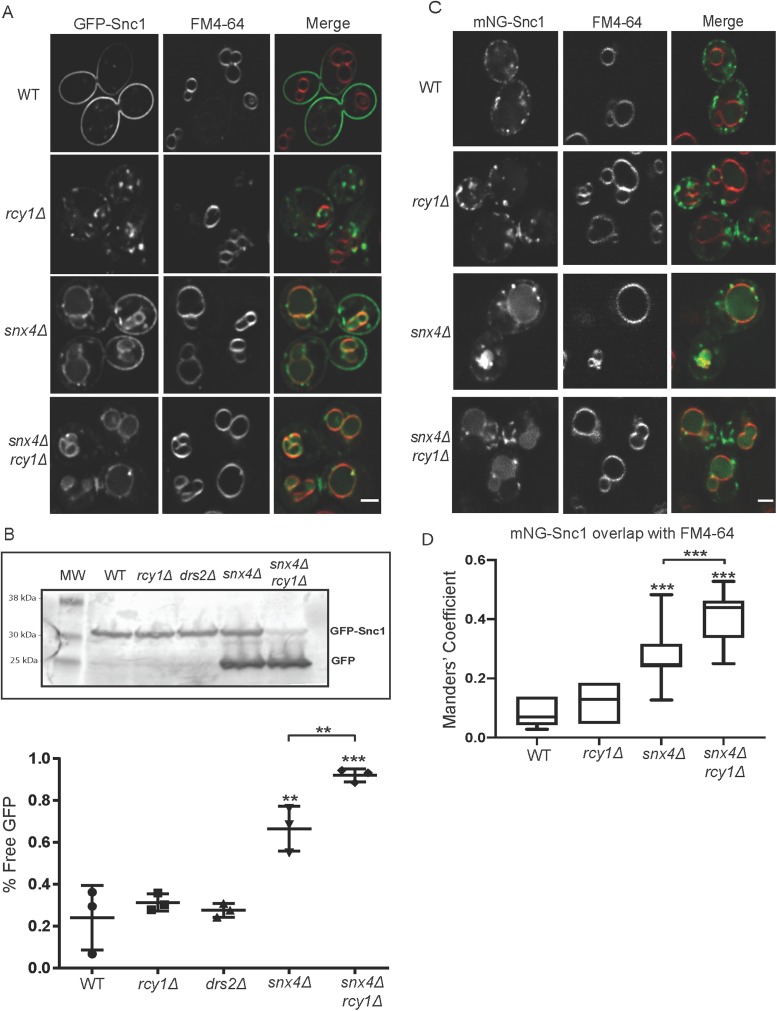
FIGURE 2:
Combined loss of Rcy1 and Snx4 enhances GFP-Snc1 missorting to the vacuole. (A) Cells expressing GFP-Snc1 were pulsed with FM4-64 and the dye was allowed to chase to the vacuole over 90 min. The cells were then imaged at 1000×; single planes are shown. (B) WT and mutant cells expressing GFP-Snc1 were cultured overnight, harvested, and lysed in buffer containing SDS. These lysates were used in a Western blot and blotted with mouse anti-GFP antibody and then a fluorescently labeled rabbit anti- mouse antibody and imaged. Lower molecular weight bands represent free GFP that has been proteolytically cleaved within the vacuole. The ratio of “free GFP” signal to total signal was used to quantify these blots. Data represent three independent experiments. (C) Cells expressing a Cu-induced mNG-Snc1 construct at endogenous level were pulsed with FM4-64, and the dye was allowed to chase to the vacuole. Micrographs were taken at 1000×; single planes are shown. (D) Channels were separated and thresholded and correlation was measured by finding the MCC between the channels (n = 50). Scale bars represent 2 µm.

