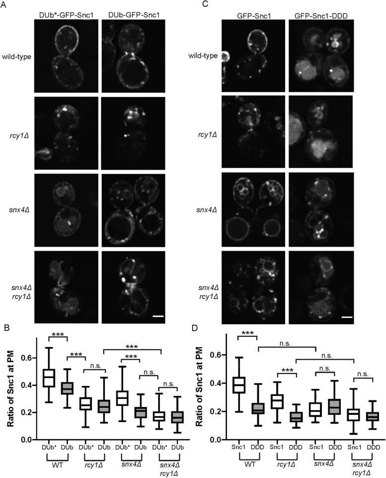
FIGURE 4:
Distinct sorting signals mediate Rcy1- and Snx4-dependent pathways. (A) WT and mutant cells expressing DUb-GFP-Snc1 and DUb*-GFP-Snc1 were imaged at 1000×; single planes are shown. (B) Quantification of images captured in A (n = 50); images were analyzed by determining the ratio of GFP signal at the PM as a function of total fluorescent signal. (C) WT and mutant cells expressing GFP-Snc1 and GFP-Snc1-DDD, both expressed from the PRC1 promoter, were imaged at 1000×; single planes are shown. The PRC1 promoter is comparable in strength to the CUP1 promoter and so approximately 40% of WT GFP-Snc1 localized to the PM in these conditions. (D) Quantification of images captured in C (n = 50); images were analyzed by determining the ratio of GFP signal at the PM as a function of total fluorescent signal.

