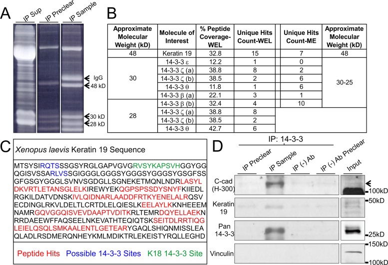
FIGURE 2:
Xenopus K19 associates with 14-3-3 proteins and C-cadherin. (A) Pan 14-3-3 immunoprecipitates (1% Tergitol type NP-40) from whole embryo lysates prior to band extraction and processing using LC/MS-MS. Prominent bands at 48, 30, and 28 kDa were processed. Heavy chain IgG from the antibody used for IP was not excised. (B) Table summary of relevant proteins detected in gel extracts processed using LC/MS-MS. Experiments were conducted using 14-3-3 immunoprecipitates from whole embryo lysates (WEL) as well as lysates from mesendoderm (ME) tissue only. Analysis was performed using Scaffold 4.7.3. (C) Summary schematic of K19 peptides (red) detected in the 48 kDa sample. Peptides are depicted within the context of the K19 primary structure and alongside described (green) and predicted (blue) possible 14-3-3 interaction sites. (D) 14-3-3 proteins were immunoprecipitated (1% Tergitol type NP-40) from whole embryo lysates and immunoblotted for C-cadherin, K19, and Vinculin. C-cadherin band is denoted by an arrow. The bottom band is yolk protein from sample.

