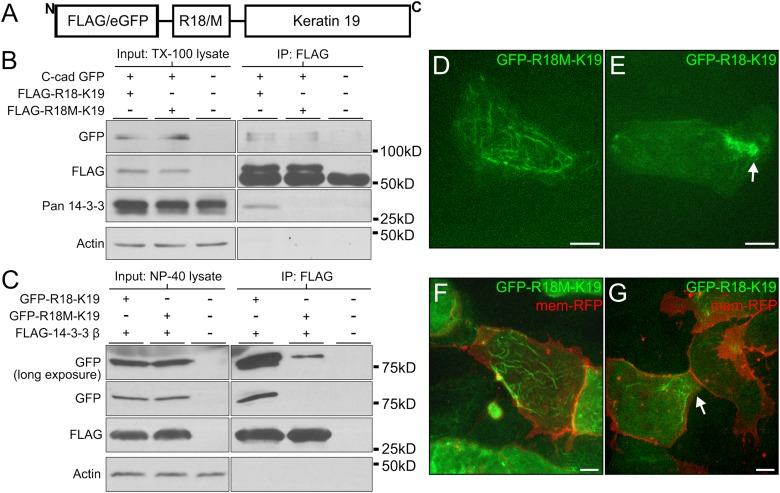
FIGURE 7:
14-3-3 proteins target keratins to cell–cell adhesions. (A) Schematic of fusion peptides created by the insertion of R18 or R18M (R18/M) into FLAG/eGFP-K19. This construct was generated using full-length K19.L from X. laevis. (B) Protein lysates from stage 10.5 Xenopus embryos expressing C-cadherin-eGFP and either FLAG-R18-K19 or FLAG-R18M-K19 were prepared in 1% Triton X-100. FLAG constructs were immunoprecipitated and analyzed by immunoblot for associated proteins. (C) Protein lysates from stage 10.5 Xenopus embryos expressing human FLAG-14-3-3β and either eGFP-R18-K19 or eGFP-R18M-K19 were prepared in 1% Tergitol-type NP-40. FLAG constructs were immunoprecipitated and analyzed by immunoblot for associated proteins. (D, E) Explanted mesendoderm cells mosaically expressing eGFP-R18M-K19 (in D) or eGFP-R18-K19 (in E). (F, G) Explanted mesendoderm coexpressing mem-RFP and eGFP-R18M-K19 (in F) or eGFP-R18-K19 (in G). Arrows indicate areas where filament densities have localized. Images are z-stacks (maximum intensity projection). Scale bars are 10 μm.

