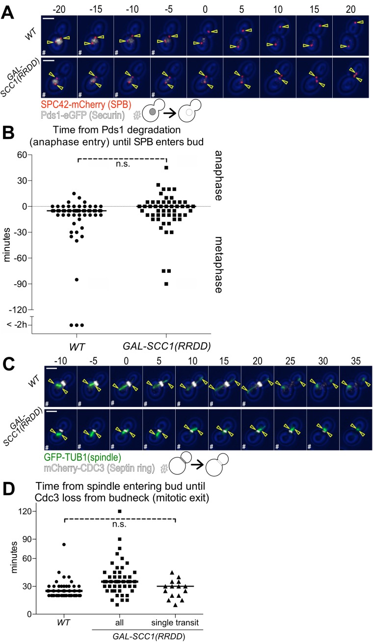
FIGURE 5:
Both SPBs moving into the daughter cell causes exit from mitosis. (A, B) Time from anaphase entry (degradation of Pds1-eGFP) until a SPB enters the bud. Wild-type (Ay40836) or GAL-SCC1(RRDD) (Ay40862) cells containing PDS1-eGFP and SPC42-mCherry were shifted to galactose containing medium at 25°C and imaged every 5 min for 8 h. GAL-SCC1(RRDD) cells were excluded if the SPBs separated (>3 μm). (A) Representative 5 min time-lapse images of a cell in metaphase degrading Pds1-eGFP, transitioning to anaphase, and translocating a SPB in the daughter cell. SPB (Spc42-mCherry) in red, Securin (Pds1-eGFP) in white, and yellow arrowheads indicate location of the SPBs determined from Spc42 localization; scale bar is 3 μm. Metaphase cells containing Pds1-eGFP are marked as “#.” (B) Time from the completion of Pds1-eGFP degradation (anaphase entry) until the first frame a SPB translocates the bud neck (n > 50 cells per condition, median displayed as a solid line, no significant difference according to Student’s t test). (C, D) Time from the spindle entering the bud until mitotic exit (mCherry-Cdc3 loss from the bud neck). Wild-type (Ay39477) or GAL-Scc1(RRDD) (Ay40483) cells containing mCherry-CDC3 and GFP-TUB1 were shifted to galactose containing medium at 25°C and imaged every 5 min for 6 h. GAL-SCC1(RRDD) cells were excluded if the SPBs separated (>3 μm). (C) Representative 5 min time-lapse images of spindle movement into the daughter cell followed by mitotic exit (mCherry-Cdc3 loss from the bud neck). Spindle (GFP-Tub1) in green, Septin ring (mCherry-Cdc3) in white, and yellow arrowheads indicate location of the SPBs determined from Tub1 localization; scale bar is 3 μm. Time points where the cell had not exited mitosis as judged by loss of Cdc3 from the bud neck are marked with “#.” (D) Time from the first frame when part of the spindle translocates the bud neck until loss of Cdc3 ring from the bud neck. The category “all” includes all cells where spindle elongation does not occur and a SPB moves into the bud at some point during imaging. The category “single transit” only encompasses cells in which the spindle transited the bud neck once and remained in the bud (WT n = 52 cells, all n = 51 cells, single transit n = 15 cells, median displayed as solid line, no significant difference according to Student’s t test between control and “single transit” cells.).

