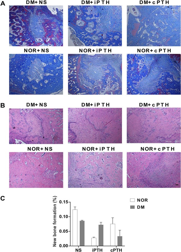
Figure 4. Histological analysis of the extraction socket. (A) Masson’s trichrome staining (red parts meant new bone and light blue parts meant collagen. Scale bar: 100μm); (B) HE staining; and (C) New alveolar bone formation in the tooth extraction socket. PTH therapy for 14 days induced no detectable new alveolar bone formation.

