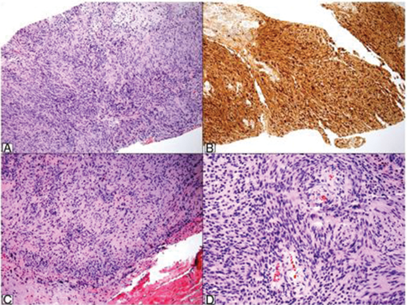
Fig. 4.

Histopathological study of biopsy specimen showing a spindle cells neoplasm with no atypia and alternating higher and lower cellularity areas (A, hematoxylin & eosin [H&E], 100×). An immunohistochemical study reveals the strong, diffuse positivity for S-100 protein (B, S-100, 100×). The surgical piece exhibited the same aspects from the biopsy, that is, a spindle cell neoplasm with short bundles, hypocellular areas and nuclear palisade foci (C and D, H&E, 100× and 200×).
