Fig. 1.
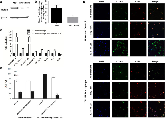
Evaluation of effects of CRISPR-RICTOR-Liposome on M2 polarized macrophages in vitro. a Immunoblotting analysis of RICTOR protein expression in untreated (MΦ) versus macrophages treated to CRISPR-CRISPR-Liposome (MΦ CRISPR). Isolated mouse macrophages were cultured, treated, and analyzed by Western blot. b Densitometric analysis of RICTOR band intensities normalized to β-actin. n = 3, *significant to untreated macrophage control (MΦ) (p < 0.05). c Effect on macrophage differentiation of CRISPR treatment targeting RICTOR, coupled with macrophage differentiation stimulated towards M1 (IFNγ + LPS) or M2 (IL-4 + M-CSF). Cells were stained with CD163 (green- M2 marker) and CD80 (red-M1 marker). d mRNA expression from M2 macrophages with and without treatment with CRISPR-RICTOR-Liposome, measured via qPCR (n = 4). e Quantitative analysis of cell phenotype. Scale bar = 100 μm, mean ± SD, biological replicates n = 5, *p < 0.05, **p < 0.01 vs. control

