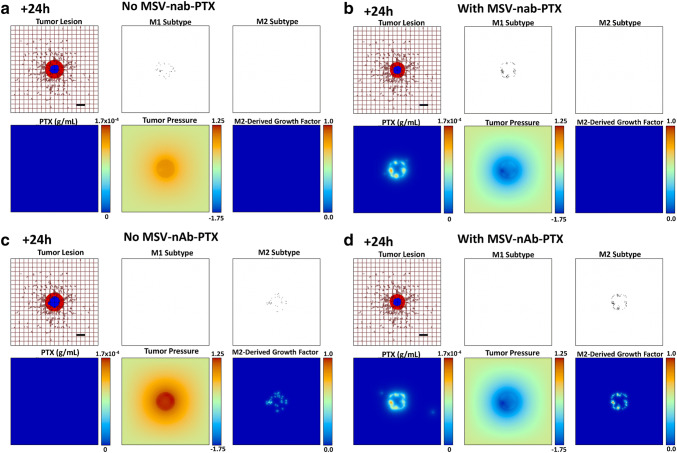
Fig. 3.
Simulation of polarized macrophage activity on a growing BCLM lesion at 24 h post-initiation. aM1-only, without MSV-nab-PTX treatment; bM1-only, with MSV-nab-PTX shown. cM2-only, without MSV-nab-PTX treatment; dM2-only, with MSV-nab-PTX. As the lesion shrinks during treatment [with viable tumor tissue (red) enclosing a hypoxic region (blue) without necrosis], the oncotic pressure (non-dimensional units) due to cell proliferation correspondingly decreases. The dense liver capillary network is modeled by the rectangular grid (brown), with irregular sprouts generated through angiogenesis during the lesion progression. The M2-derived growth factor (non-dimensional units) is only present for the M2 case. Bar = 200 μm

