Figure 3.
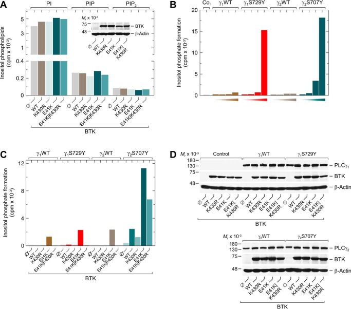
Activation of PLCγ2S707Y by catalytically-inert BTK is not mediated by augmented PLC substrate availability. A, COS-7 cells were transfected as indicated with 2 μg/well of empty vector or 0.5 μg/well of vector encoding WT BTK (WT) or BTK variants K430R, E41K, or E41K/K430R. Twenty four hours after transfection, the cells were incubated for 18 h with myo-[2-3H]inositol. Then, the amounts of radioactively labeled PtdIns (PI), PtdInsP (PIP), and PtdInsP2 (PIP2) in the intact COS-7 cells were determined by TLC analysis as described under “Experimental procedures.” Inset, cells from one well each were lysed and subjected to SDS-PAGE. Subsequent immunoblotting was performed using an antibody reactive against BTK or antibody reactive against β-actin. B, COS-7 cells were transfected as indicated with 500 ng of empty vector (Co.) or increasing amounts (15, 50, 150, or 500 ng) of vector encoding WT PLCγ1 (γ1WT), PLCγ1S729Y (γ1S729Y), WT PLCγ2 (γ2WT), or PLCγ2S707Y (γ2S707Y). C, COS-7 cells were transfected as indicated with 50 ng/well of vectors as described in B, and 100 ng/well of vector encoding WT BTK (WT) or BTK variants K430R, E41K, or E41K/K430R. Analysis of inositol phosphate formation was done as in Fig. 1. D, cells from one well each functionally analyzed in C were washed with 0.2 ml of Dulbecco's PBS and then lysed by addition of 100 μl of SDS-PAGE sample preparation buffer. Aliquots of the samples were subjected to SDS-PAGE, and immunoblotting was performed using an antibody reactive against the c-Myc epitope present on WT PLCγ1, WT PLCγ2, PLCγ1S729Y, and PLCγ2S707Y, antibody reactive against BTK, or antibody reactive against β-actin. A–C show representative results from three independent experiments each as mean values of three technical replicates.

