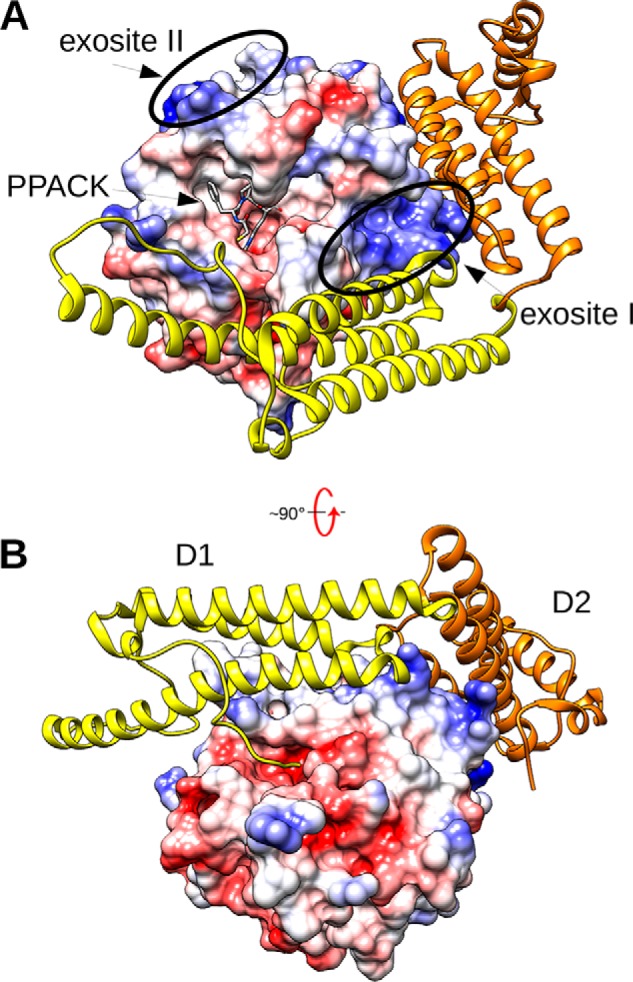
Figure 2.

The SC(1–325)·Pre2 complex. A, the molecular surface of Pre2 is shown colored by electrostatic potential, with the ATA-PPACK (N-(sulfanylacetyl)-d-phenylalanyl-N-[(2S,3S)-6-{[amino(iminio)methyl]amino}-1-chloro-2-hydroxyhexan-3-yl]-l-prolinamide) inhibitor displayed as gray sticks in the active site. SC(1–325) is displayed in ribbon mode, with the N-terminal D1 domain colored yellow and the D2 domain colored gold. B, the complex above is rotated ∼90º from the standard orientation to show the insertion of the N-terminal SC peptide Ile1-Val2-Thr3 into the Ile16-binding pocket of Pre2, triggering activation. This figure was constructed with UCSF Chimera using the X-ray crystal structure 1NU9.pdb (16).
