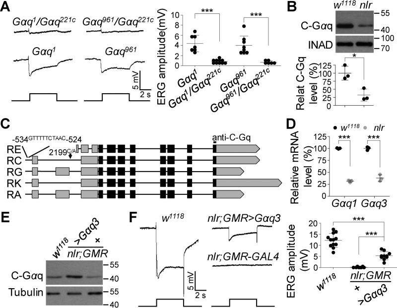
Figure 4.
Gαq3 isoform contributes to the residual photoresponse in Gαq1 isoform null mutants. A, representative ERG traces of Gαq1/Gαq221c and Gαq961/Gαq221c flies. For all ERG traces, event markers represent 5-s light pulses, and scale bars are 5 mV. Quantification of ERG amplitudes for each genotype is presented in the right panel. B, Western blots of Gαq3 protein levels in w1118 and nlr mutant photoreceptor cells. The eye-specific protein INAD was used as a loading control. Quantification of Gαq3 levels for each genotype is presented in the lower panel. C, additional mutations in the Gαq gene. The point mutation in the nlr allele is marked with a black arrow, and the sequence inserted upstream of the transcription start site is listed at the top. The anti-C-Gαq antibody-recognized site is labeled at the top. D, mRNA levels of Gαq1 and Gαq3 in w1118 and nlr mutant retina. Total RNA was extracted from isolated adult retina. Rp49 was used as an internal control. Data are presented as mean ± S.D. from three independent experiments. E, Western blots show Gαq3 protein levels in rescued flies. F, representative ERG traces of w1118, nlr;GMR-GAL4, and nlr;GMR-GAL4/UAS::Gαq3 flies. For all ERG traces, event markers represent 5-s light pulses, and scale bars are 5 mV. Quantification of ERG amplitudes for each genotype is presented in the right panel.

