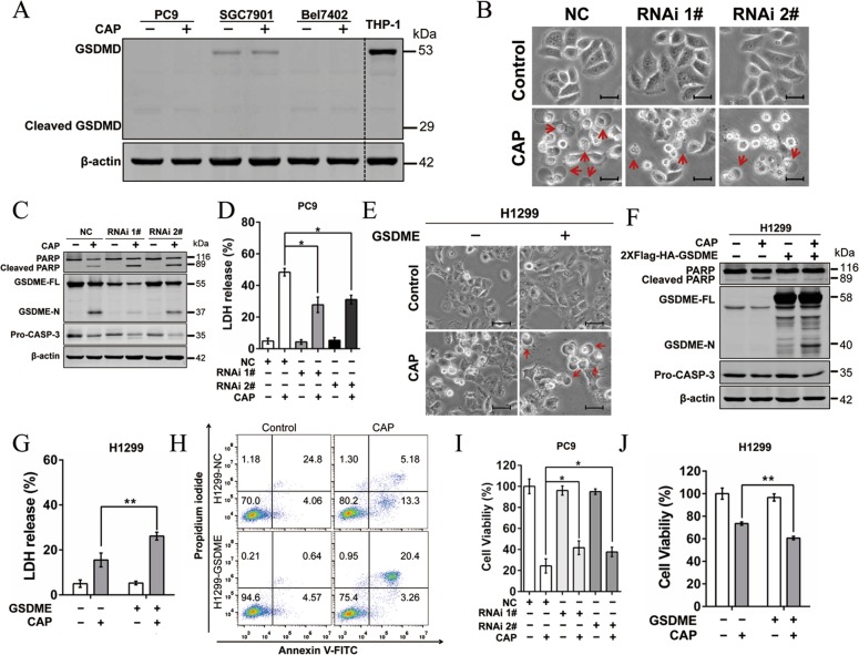
Fig. 3. GSDME is essential in CAP-induced pyroptosis.
a GSDMD-FL and cleaved GSDMD (GSDMD-N) were detected by western blotting in PC9, SGC-7901 and Bel7402 at 24 h after CAP exposures. THP1 cells expressing GSDMD were used as a positive control. b–d Alterations in features of cell pyroptosis were determined upon CAP treatment after knocking down GSDME in PC9 cells. b Representative microscopic images of negative control (NC) and GSDME-knockdown PC9 cells (RNAi#1 and RNAi#2). Red arrowheads indicated large bubbles emerging from the plasma membrane. Scale bar, 25 µm. c The apoptosis- and pyroptosis-related proteins including PARP, cleaved-PARP, GSDME, GSDME-N and pro-CASP-3 analyzed by western blotting. β-actin served as loading control. d LDH release in the culture supernatants. e–g The features of cell pyroptosis were determined upon CAP treatment after overexpressing GSDME in H1299 cells. e Representative microscopic images. Red arrowheads indicated large bubbles emerging from the plasma membrane. Scale bar, 25 µm. f Apoptosis- and pyroptosis-related proteins including PARP, cleaved-PARP, GSDME, GSDME-N and Pro-CASP-3 determined by western blotting. g LDH release assays. h Annexin V-FITC/PI assay was performed to identify the pyroptotic and apoptotic cells after CAP treatment in H1299 and H1299-GSDME cells. i, j Cell viability was measured at 24 h after CAP exposures in GSDME knockdown PC9 cells (i) and GSDME overexpressed H1299 cells (j), respectively. All the data are presented as the mean ± SD from three independent experiments. *p < 0.05.

