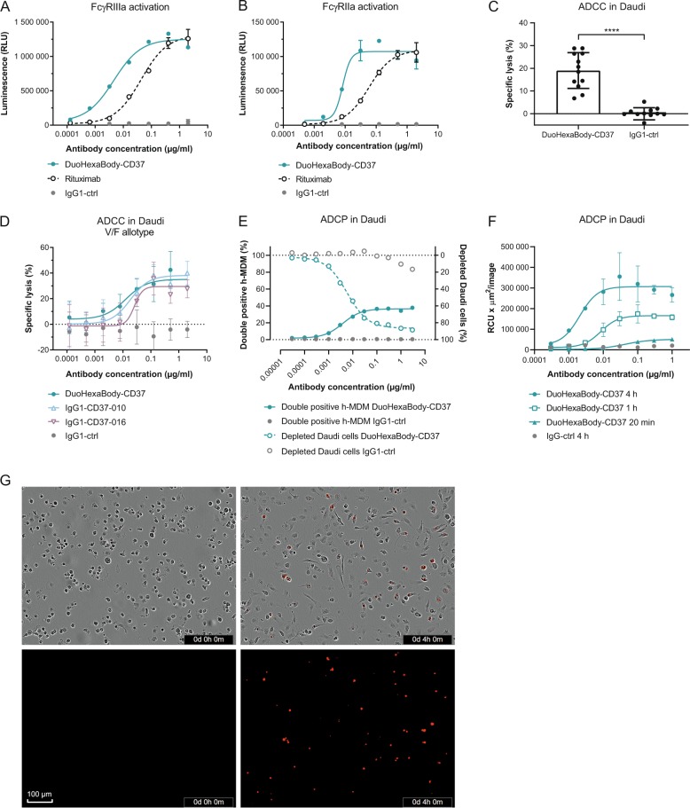
Fig. 4. DuoHexaBody-CD37 induces efficient ADCC and ADCP in vitro.
a, b FcγRIIIa (a) and FcγRIIa (b) crosslinking by DuoHexaBody-CD37 was analyzed in a bioluminescent Reporter Bioassay using Daudi target cells and engineered FcγRIIIa- or FcγRIIa-expressing Jurkat effector T cells that express luciferase upon FcγR crosslinking. Luciferase production is presented as relative luminescence units (RLU). Error bars represent the mean ± SD of duplicate measurements. c ADCC by 2 µg/ml DuoHexaBody-CD37 was evaluated in a classical 51Cr release assay using Daudi target cells and PBMCs from 12 healthy human donors as a source of effector cells (E:T of 100:1). The percentage lysis was calculated relative to a Triton X-100 control (100% lysis) and no antibody control (0% lysis). ****P < 0.00001, paired T-test with two-tailed 95% confidence intervals. d Dose-response ADCC (mean percentage lysis ± SD of three replicate samples) induced by DuoHexaBody-CD37, WT IgG1-CD37-010 and WT IgG1-CD37-016 shown for one representative responsive donor as described in c. Error bars represent the mean ± SD of triplicate measurements. e ADCP induced by DuoHexaBody-CD37 using Daudi target cells and monocyte-derived h-MDM from healthy human donors as a source of effector cells. Calcein AM-labeled Daudi cells opsonized with DuoHexaBody-CD37 were incubated with CD11b + h-MDM at an E:T ratio of 2:1 and ADCP was analyzed by flow cytometry after a 4 h co-culture. The amount of h-MDMs that phagocytosed Daudi cells is presented as percent CD11b+/calcein AM+/CD19- double positive cells. CD19 was used to exclude macrophages with bound instead of phagocytosed tumor cells. The percentage CD11b−/calcein AM+ cells was determined as an indicator of the amount of non-phagocytosed Daudi cells; presented here as a depleted cell fraction relative to a no antibody control sample. Data from one representative donor out of three is shown. f ADCP of pHRodo-labeled Daudi target cells by h-MDM induced by DuoHexaBody-CD37 over time at an E:T ratio of 1:1, shown for one out of three representative donors. Red fluoresence indicates phagocytosed Daudi target cells by h-MDM. ADCP was quantified by the total sum of the red fluorescent intensity in the image (RCUxµm2/image) and presented as the mean ± SD of duplicate measurements. g Phase contrast- and red fluorescent images of DuoHexaBody-CD37-opsonized (1 µg/ml) pHRodo-labeled Daudi target cells co-cultured with h-MDM effector cells at 0 and 4 h incubation as described in f.

