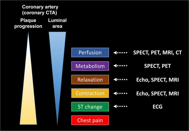
Fig. 1.

Illustration showing the progressive pathological conditions in the myocardial ischemic cascade. Coronary artery atherosclerosis progresses and leads to myocardial hypoperfusion because of plaque progression and luminal stenosis. Myocardial hypoperfusion is followed by metabolic abnormalities, diastolic dysfunction, systolic dysfunction, and ECG abnormalities, culminating in chest pain. The right column shows the modalities that can be used to detect abnormalities at each step of the cascade. CT computed tomography, CTA computed tomography angiography, ECG electrocardiogram, SPECT single-photon emission computed tomography, PET positron emission tomography, MRI magnetic resonance imaging
