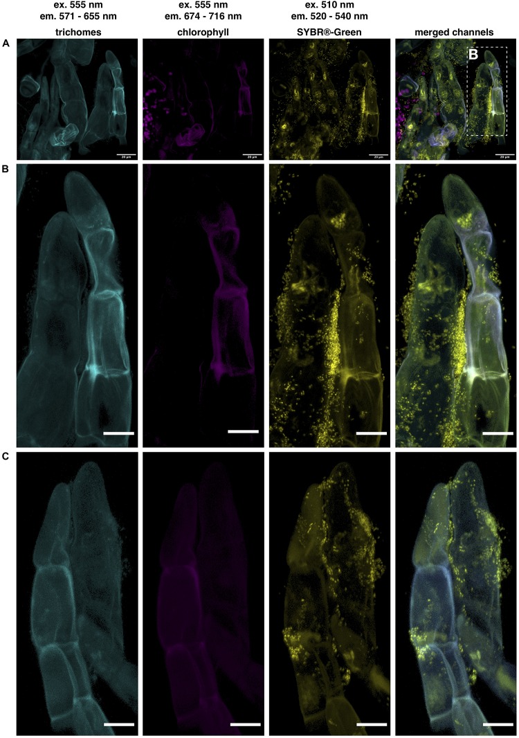
FIGURE 2.
Microscopic analysis of bacteria associated to secretory trichomes on the D. bulbifera leaf acumen. Confocal laser scanning microscopy (CLSM) images of trichomes located on the upper side of the leaf acumen. Trichome autofluorescence could be separated from other channels and can be observed in cyan, chlorophyll autofluorescence in magenta and nucleic acids were stained with SYBR® < /cps:sup > -Green, here depicted in yellow. Excitation (ex.) and emission (em.) wavelengths are indicated above. (A) Trichomes densely cover the surface of the acumen and microorganisms strongly colocalize with trichomes, as can be observed in panel (B) a magnification of two trichomes in panel (A), as well as in panel (C), showing another pair of trichomes densely colonized by microorganisms. Scale bars represent 20 and 10 μm in panels (A) and (B,C), respectively.

