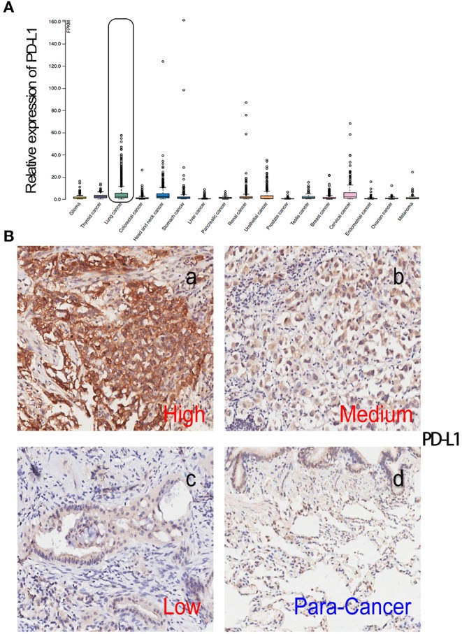
Figure 2.

The expression level of PD-L1 in patients with NSCLC. (A) The expression level of PD-L1 in all types of cancer, data were from the Protein Atlas website: https://www.proteinatlas.org/ENSG00000120217-CD274/pathology; (B) lung tumor tissue and para-cancer lung tissue were stained with PD-L1 antibody. Using both the IHC staining intensity and proportion of cell stained, the IHC score was determined by a semi-quantitative method. The expression level was further classified into low expression (≤1), moderate expression (1.5-6) and strong positive expression (≥7.5) for PD-L1. A reprehensive image of high-, medium- and low-level of PD-L1 expression in tumor tissue from 180 patients with NSCLC receiving pulmonectomy (a, b, c), and PD-L1 expression in its corresponding para-cancer tissue (d).
