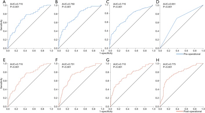
Figure 3.
The ROC curves of predictive models. (A) The ROC curve of the model for LNM with the preoperative independent risk factors; (B) the ROC curve of the model for LVI with the preoperative independent risk factors; (C) the ROC curve of the model for SM invasion with the preoperative independent risk factors; (D) the ROC curve of the model for MM invasion with the preoperative independent risk factors; (E) the ROC curve of the model for LNM with the postoperative independent risk factors screened out from the variates without depth of invasion and LVI; (F) the ROC curve of the model for LNM with the postoperative independent risk factors screened out from the variates containing depth of invasion; (G) the ROC curve of the model for LNM with the postoperative risk factors screened out from the variates containing LVI; (H) the ROC curve of the model for LNM with the postoperative risk factors screened out from the variates containing depth of invasion and LVI. AUC, areas under the ROC curve; LNM, lymph node metastasis; LVI, lymphovascular invasion; MM, muscularis mucosa.

