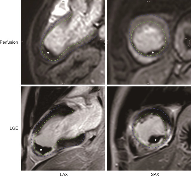
Figure 2.
Manifestations of left ventricular thrombus (LVT) on CMR. LVT (white star) appears as a hypo-signal mass in the left ventricular in both the gadolinium perfusion (upper row) and late enhancement (lower row) imaging. Endo- and epi-cardium is delineaed by green and blue dash line, respectively. LGE, late gadolinium enhancement; LAX, long axis; SAX, short axis.

