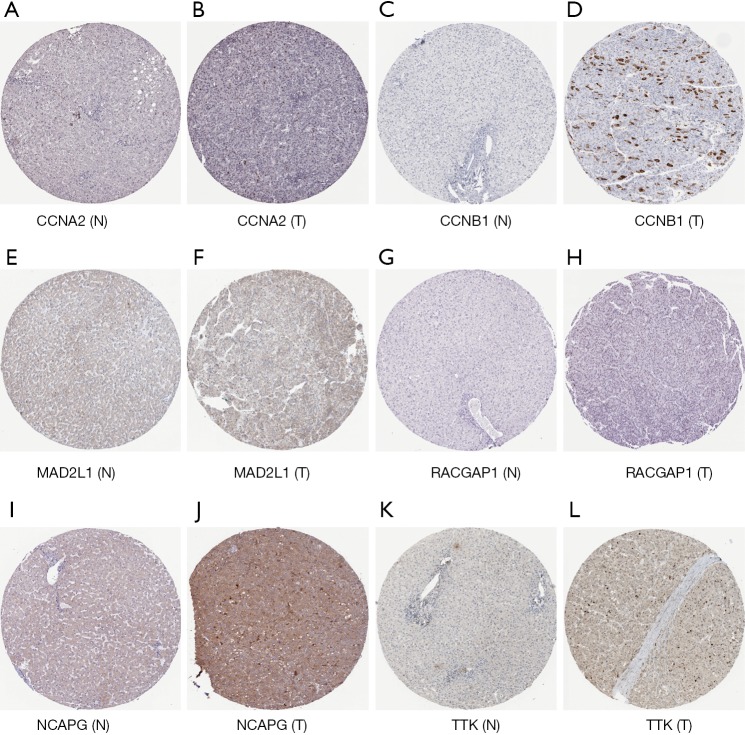
Figure 7.
Protein expression of CCNA2, CCNB1, MAD2L1, RACGAP1, NCAPG, TTK. The tissue microarray using paired tumor tissue and adjacent normal tissue from the margin of the tumor. Immunohistochemistry of tissue microarray was performed by using the antibody (CAB000114, CAB003804, HPA003348, HPA043912, HPA039613, CAB013229, Abcam). (A) Protein expression of CCNA2 in normal tissue (not detected); (B) protein expression of CCNA2 in tumor tissue (staining: medium; intensity: strong; quantity: <25%); (C) protein expression of CCNB1 in normal tissue (not detected); (D) protein expression of CCNB1 in tumor tissue (staining: medium; intensity: strong; quantity: <25%); (E) protein expression of MAD2L1 in normal tissue (not detected); (F) protein expression of MAD2L1 in tumor tissue (staining: low; intensity: weak; quantity: 75–25%); (G) protein expression of RACGAP1 in normal tissue (not detected); (H) protein expression of RACGAP1 in tumor tissue (staining: medium; intensity: moderate; quantity: 75–25%); (I) protein expression of NCAPG in normal tissue (not detected); (J) protein expression of NCAPG in tumor tissue (staining: medium; intensity: moderate; quantity: >75%); (K) protein expression of TTK in normal tissue(not detected); (L) protein expression of TTK in tumor tissue (staining: low; intensity: moderate; quantity: <25%).

