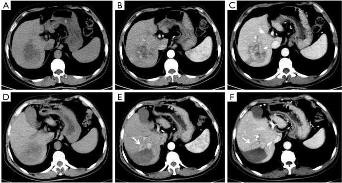
Figure 2.
A 70-year-old man with hepatocellular carcinoma (HCC) in the VII segment of the liver. A tumor 6.9 cm in size in the right lobe of liver with nonuniform low-density (A), showing hyperenhancement in the arterial phase (B), and continuous enhancement without distinct “washout” appearance in the portal venous phase (C); after radiofrequency ablation (RFA), a non-enhanced ablation area was found in original lesion area, along with a 5.0 cm-sized irregular, thickened tissue area (arrows) along the margin of the treated lesion (D), showing hyperenhancement in the arterial phase (E) and washout (F). Diagnosis was agreed upon by the 2 readers (LR-TR viable 5.0 cm).

