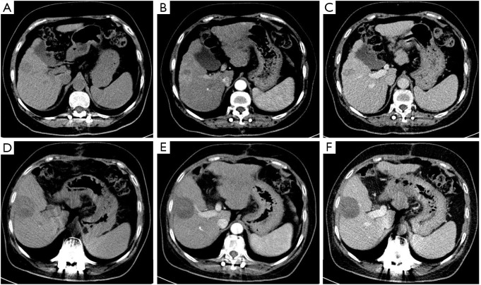
Figure 3.
A 72-year-old woman with hepatocellular carcinoma (HCC) in the V segment of the liver. A low-density tumor 2.5 cm in size on normal abdominal CT images (A), showing hyperenhancement (rim) in the arterial phase (B), “washout” appearance, and enhanced capsule (arrow) in the portal venous phase (C); after radiofrequency ablation (RFA), a non-enhanced ablation area was shown (D), and there was not any enhancement tissue in or along the margin of the treated lesion (E,F). Diagnosis was agreed upon by the 2 readers (LR-TR nonviable).

