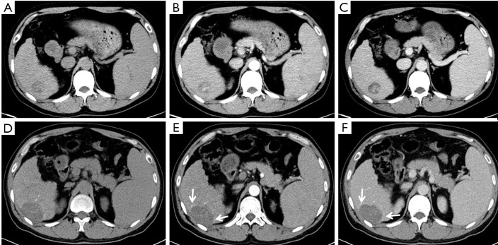
Figure 4.
A 48-year-old man with hepatocellular carcinoma (HCC) in the VI segment of the liver. A low-density lesion in the right lobe of the liver 2.7 cm in size on normal abdominal CT images (A) with nonuniform hyperenhancement (not rim) in the arterial phase (B) and “washout” appearance in the portal venous phase (C); after radiofrequency ablation (RFA), a non-enhanced ablation area was shown (D), but with no nodular or irregular enhancement except for an nonsmoothed rim (arrows) of enhancement on arterial (E) and portal venous phases (F). There was inconsistent diagnosis between the 2 readers (R1: equivocal; R2: nonviable. The final conclusion re-valuated by R3: equivocal).

